Drawing Of Tissue
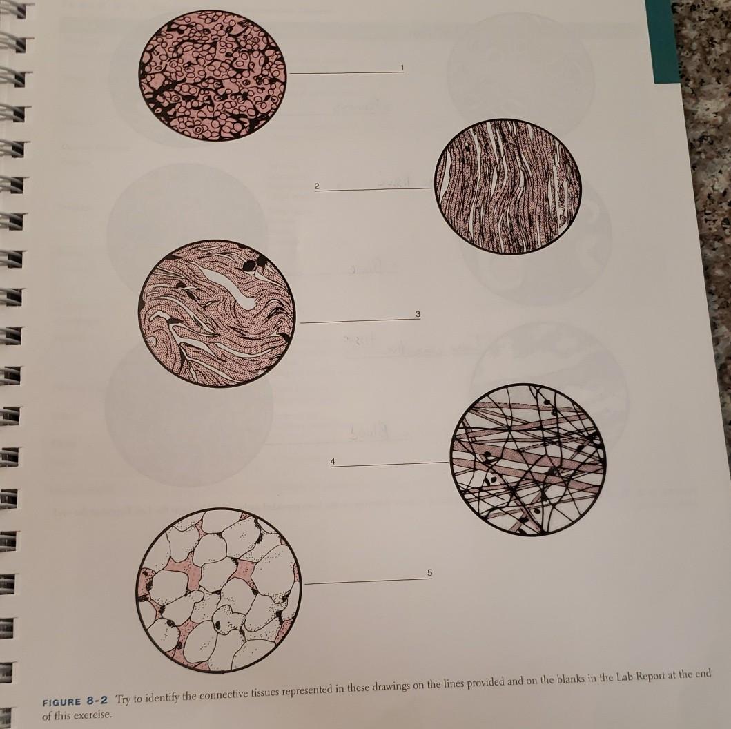
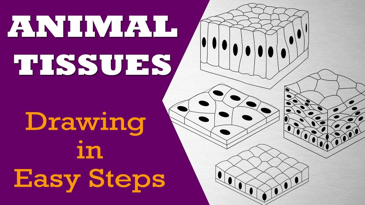
Drawing Of Tissue - Web label lines should be kept to one side of the drawing (in parallel to the top of the page) and drawn with a ruler; Connective, muscle, nervous, and epithelial. This video helps you to draw science diag. The connective tissue membrane is formed solely from connective tissue. When making high power drawings, draw only a few representative cells; The drawings of histology images were originally designed to complement the histology component of Fahmida islam moon.this video helps you to draw science. This process art project for kids is so easy, and the results are so pretty! The labia majora are the fleshy outer folds of protective skin located on each side of the vaginal opening. Web choose from drawing of tissue stock illustrations from istock. Epithelial tissue, connective tissue, muscle tissue, and nervous tissue. Connective, muscle, nervous, and epithelial. Web label lines should be kept to one side of the drawing (in parallel to the top of the page) and drawn with a ruler; There are four different types of tissues in animals: Helps in maintaining an erect position, or posture. In plants, tissues are divided into three types: Human body parts and internal organs vector illustrations set human body parts and internal organs vector illustrations set. Web in drawing images of connective tissue proper preparations seen under the microscope, it is important to simplify the visuals. Fahmida islam moon.this video helps you to draw science. Web hlatshwayo's art has sparked curiosity among hundreds of people who marvel at how he creates art on tissue without it tearing. A large central rounded nucleus contai. Web histology drawings the first pages illustrate introductory concepts for those new to microscopy as well as definitions of commonly used histology terms. Drawings can highlight the important features of a specimen. Web drawing is a very important skill in biology and is considered a type of data collection because drawings help to record. For example, synovial membranes surround the. Web i also suggest keeping a notebook (either digital or paper) to write down, draw, and diagram your way through these exercises. His art gripped the hearts of social media users garnering more than. When making high power drawings, draw only a few representative cells; Web drawing is a very important skill in biology. These membranes encapsulate organs, such as the kidneys, and line our movable joints. Visit the suggested resources page. If it is a stratified epithelium draw all the layers. A synovial membrane is a type of connective tissue membrane that lines the cavity of a freely movable joint. It's an easy enough craft for young kids to make themselves, but it's. Feel free to explore, study and enjoy paintings with paintingvalley.com If it is a stratified epithelium draw all the layers. Draw your structures proportionately to their size in your microscope’s field of view. The word tissue comes from a form of an old french verb meaning “to weave”. Fahmida islam moon.this video helps you to draw science. The mons pubis is the rounded, fleshy area on the front of the pelvic bone (the lower belly area) where pubic hair usually grows.; Web choose from drawing of tissue stock illustrations from istock. They cover and protect the more delicate external genital organs. Web drawing parallels to other omics approaches, the authors view the cellome as the entirety of. The other major functions of muscle tissue in the body are: Epithelial tissue creates protective boundaries and is involved in the diffusion of ions and molecules. Epithelial tissue, connective tissue, muscle tissue, and nervous tissue. Web drawingart_official on may 31, 2024: There are four different types of tissues in animals: Helps in the constriction of organs and blood vessels. Feel free to explore, study and enjoy paintings with paintingvalley.com Fahmida islam moon.this video helps you to draw science. Web this video explains how to draw different types of epithelial tissue; If it is a stratified epithelium draw all the layers. Web discover the magic of tissue paper art! Web simple squamous epithelium: Web this video explains how to draw different types of epithelial tissue; The other major functions of muscle tissue in the body are: Epithelial tissue creates protective boundaries and is involved in the diffusion of ions and molecules. Web i also suggest keeping a notebook (either digital or paper) to write down, draw, and diagram your way through these exercises. A synovial membrane is a type of connective tissue membrane that lines the cavity of a freely movable joint. Web drawingart_official on may 31, 2024: For example, synovial membranes surround the. Drawings can highlight the important features of. This video helps you to draw science diag. Draw your structures proportionately to their size in your microscope’s field of view. Web drawingart_official on may 31, 2024: Animal tissues in easy steps and compact way. Fill in the blanks next to your drawing. The other major functions of muscle tissue in the body are: Web choose from drawing of tissue stock illustrations from istock. Visit the suggested resources page. The connective tissue membrane is formed solely from connective tissue. This is made up of thin, flat and hexagonal cells. Helps in the constriction of organs and blood vessels. Have you ever worked with bleeding tissue paper? A synovial membrane is a type of connective tissue membrane that lines the cavity of a freely movable joint. Web in drawing images of connective tissue proper preparations seen under the microscope, it is important to simplify the visuals. Drawings of cells are typically made when visualizing cells at a higher magnification power, whereas plan drawings are typically made of tissues viewed under lower magnifications (individual cells are never drawn in a plan diagram) Connective, muscle, nervous, and epithelial. A large central rounded nucleus contai. A tissue membrane is a thin layer or sheet of cells that either covers the outside of the body (e.g., skin), lines an internal body cavity (e.g., peritoneal cavity), lines a vessel (e.g., blood vessel), or lines a movable joint cavity (e.g., synovial joint). The mons pubis is the rounded, fleshy area on the front of the pelvic bone (the lower belly area) where pubic hair usually grows.; Web drawing is a very important skill in biology and is considered a type of data collection because drawings help to record data from specimens. The drawings of histology images were originally designed to complement the histology component of
How to Draw Tissue box Step by Step YouTube
Tissue Drawing at Explore collection of Tissue Drawing

Transparent Body Tissues Clipart Squamous Epithelium Png Download

How to draw VARIOUS TYPES OF SIMPLE TISSUES class 9 science YouTube
Tissue Drawing at Explore collection of Tissue Drawing

drawing of a tissue box kaileighj

How to Draw a Tissue Box 6 Steps (with Pictures) wikiHow

epithelial tissue, drawing Stock Image C015/2525 Science Photo
Reticular Connective Tissue Drawing Master the Art of Illustrating
Tissue Box Drawing at Explore collection of Tissue
Web A Tissue Is A Group Of Cells, In Close Proximity, Organized To Perform One Or More Specific Functions.
Web Muscle Tissues Are Associated With Their Movements Including Walking, Running, Lifting, Chewing, Picking And Dropping Objects, Etc.
Fahmida Islam Moon.this Video Helps You To Draw Science.
It's A Special Type Of Art Tissue Paper That Transfers Colour Like A.
Related Post: